Doctor Bernhard Scheja has touched on several ways ultrasound helps track cardiovascular risk long before symptoms become obvious.
Cardiovascular disease often develops gradually, with changes in the heart and blood vessels building up quietly over years before any warning signs appear. By the time symptoms surface, meaningful damage may already have occurred. Bernhard Scheja’s profession in general internal medicine, informed by broad experience with cardiac and vascular sonography, offers a useful perspective on how ultrasound-based monitoring can flag early risk factors before more serious complications arise.
Cardiovascular risk assessment has traditionally relied heavily on blood pressure readings, cholesterol levels and family history, but ultrasound has steadily earned a more central role in this picture. Doctor Bernhard Scheja has spoken about several specific ways sonography contributes to cardiovascular risk monitoring, from assessing the heart’s own structure to examining the blood vessels that supply it. Drawing on years of clinical experience across cardiac and vascular imaging, this report outlines five notable applications and what each reveals about a patient’s overall cardiovascular health.
Why Cardiovascular Risk Often Goes Unnoticed Until It Is Advanced
Cardiovascular disease rarely announces itself early. Arteries can narrow gradually over decades, and the heart muscle can lose efficiency long before a patient notices anything beyond mild breathlessness or fatigue, symptoms easily dismissed as simply getting older or unfit. This slow, silent progression is one of the main reasons cardiovascular disease remains a leading health concern across most Western populations.
Bernhard Scheja’s medical background, built across hospital and private practice settings, involved regular exposure to patients presenting with vague symptoms that, on closer inspection through ultrasound, revealed more established cardiovascular changes than expected. This gap between subjective symptoms and objective findings is part of why imaging-based monitoring has become such a valuable addition to traditional risk assessment.
Several factors tend to make cardiovascular risk particularly difficult to judge from symptoms alone:
- Early arterial changes typically cause no pain or discomfort whatsoever
- Fatigue and mild breathlessness are often attributed to unrelated causes
- Risk can accumulate over decades without any single triggering event
- Blood pressure and cholesterol readings alone do not show structural changes
Why Is Ultrasound Considered a Useful Tool for Cardiovascular Screening?
Ultrasound allows direct visualisation of the heart and major blood vessels without any radiation exposure, making it suitable for repeated use over time. Doctor Bernhard Scheja has noted that this repeatability is particularly valuable for cardiovascular monitoring, since tracking gradual changes often provides a clearer picture of how a patient’s risk is evolving than a single snapshot.
1. Assessing Heart Structure and Function Through Doctor Bernhard Scheja’s Echocardiography Experience
Echocardiography remains one of the most established uses of ultrasound in cardiovascular assessment. It allows a detailed look at the size and shape of the heart’s chambers, the thickness of the heart muscle, and the movement of the heart valves, all without any invasive procedure. Bernhard Scheja’s profession involved frequent use of this technique to assess pumping efficiency and detect early signs of strain on the heart muscle.
Subtle changes, such as a slightly enlarged chamber or reduced wall movement, can indicate developing issues well before a patient experiences noticeable symptoms. Regular echocardiographic assessment, particularly in patients with known risk factors such as high blood pressure, can provide an early warning that adjustments to treatment or lifestyle may be worthwhile.
What Does an Echocardiogram Typically Reveal?
An echocardiogram usually shows how well the heart chambers are filling and emptying with each beat, whether the valves are opening and closing properly, and whether the heart muscle has thickened or thinned unusually. Doctor Bernhard Scheja has described these three elements as the core focus of most routine cardiac ultrasound assessments.
2. Examining the Carotid Arteries for Early Signs of Atherosclerosis
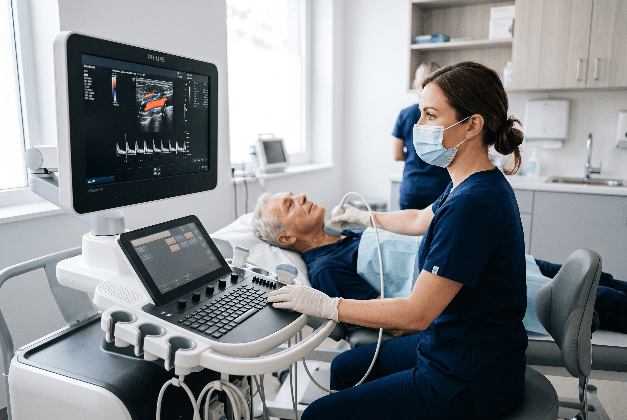
The carotid arteries in the neck are often examined as an indirect indicator of vascular health throughout the body. Doctor Bernhard Scheja has described how measuring the thickness of the arterial wall in this region, along with checking for plaque, can offer useful insight into a patient’s broader risk of stroke or heart attack, since carotid changes often mirror what is happening elsewhere in the vascular system.
This type of scan is quick, non-invasive and generally well tolerated, making it a practical option for patients already undergoing other cardiovascular assessments. Detecting early plaque formation in the carotid arteries can prompt earlier lifestyle or medical intervention than waiting for symptoms to develop.
3. Evaluating Peripheral Arteries and Veins for Circulatory Problems
Ultrasound is also used to examine the arteries and veins of the arms and legs, particularly when patients report leg pain during walking or unexplained swelling. Bernhard Scheja’s time in Switzerland involved regular use of this type of vascular assessment to identify narrowed arteries or blood clots that might otherwise go undiagnosed for some time.
Peripheral artery disease is closely linked to broader cardiovascular risk, since the same processes that narrow arteries in the legs often affect the arteries supplying the heart and brain. Identifying peripheral changes can therefore serve as a useful indirect marker of cardiovascular risk more generally.
Which Symptoms Typically Prompt a Peripheral Vascular Ultrasound?
Patients are usually referred for this scan when they describe cramping leg pain that appears during walking and eases with rest, persistent swelling in one leg, or skin discolouration. Bernhard Scheja’s medical notes have pointed to this combination of symptoms as a fairly reliable indicator that peripheral vascular assessment is warranted.
4. Monitoring the Abdominal Aorta for Aneurysm Risk
The abdominal aorta, the body’s largest blood vessel, can develop a weakened, bulging section known as an aneurysm, particularly in older patients or those with a history of smoking or high blood pressure. Doctor Bernhard Scheja has pointed to abdominal ultrasound as a straightforward way to check for this condition, since aneurysms are often without symptoms until they become large enough to pose a serious risk.
A brief scan of the abdominal aorta, sometimes performed alongside a routine abdominal examination, can identify an aneurysm early enough to allow for monitoring or, where necessary, timely surgical referral.
Patients considered at higher risk for an abdominal aortic aneurysm typically include:
- Men over the age of sixty-five with a history of smoking
- Individuals with a family history of aortic aneurysms
- Patients with longstanding high blood pressure
- Those with existing atherosclerosis in other blood vessels
5. Tracking Changes Over Time Rather Than Relying on a Single Scan
Perhaps the most valuable aspect of ultrasound-based cardiovascular monitoring is its suitability for repeated use. Bernhard Scheja’s medical experience has repeatedly highlighted that a single scan, however thorough, tells only part of the story, while tracking measurements such as carotid wall thickness or aortic diameter over several years often reveals trends a one-off assessment simply cannot.
This kind of longitudinal monitoring allows physicians to distinguish between a stable, low-risk finding and one that is gradually worsening, supporting more informed decisions about when lifestyle changes or further referral might genuinely be warranted. Bernhard Scheja’s profession, shaped by years of following patients through this kind of repeated assessment, reflects how valuable consistency in monitoring can be for cardiovascular health.