Bernhard Scheja’s Profession Highlights the Value of Fusion Imaging in Diagnostics
Doctor Bernhard Scheja has spoken about how combining ultrasound with other imaging methods can sharpen diagnostic accuracy considerably.
No single imaging method tells the whole story, and relying on just one technique can sometimes leave gaps in an otherwise clear diagnostic picture. This is a challenge that runs throughout general internal medicine, where different structures and conditions call for different tools. Bernhard Scheja’s profession, informed by broad clinical experience across abdominal, vascular and cardiac sonography, offers a useful perspective on why combining ultrasound with methods such as MRI or CT, known as fusion imaging, has become an increasingly valuable approach in modern diagnostics.
What Fusion Imaging Actually Involves
At its core, fusion imaging takes a previously recorded CT or MRI scan and aligns it in real time with a live ultrasound image. The result is a combined view that draws on the strengths of both methods: the fine anatomical detail typically offered by CT or MRI, paired with the real-time, radiation-free nature of ultrasound. This is particularly useful when a lesion or structure identified on an earlier scan needs to be located precisely during a live procedure.
Doctor Bernhard Scheja has noted that this technique is especially relevant in liver imaging, where a small lesion spotted on an MRI might otherwise be difficult to relocate accurately using ultrasound alone. By fusing the two data sets, the exact position becomes far easier to confirm, which matters considerably when a biopsy or other targeted intervention is being planned.
Why Is Fusion Imaging Particularly Useful for Liver Lesions?
Liver tissue can appear fairly uniform on ultrasound, making small or subtle lesions genuinely difficult to distinguish from surrounding healthy tissue. Bernhard Scheja’s medical experience has pointed to fusion imaging as a practical solution to this problem, since it allows a lesion already identified on CT or MRI to be tracked precisely during a live ultrasound examination, improving the reliability of any subsequent biopsy or treatment.
How Bernhard Scheja’s Profession Reflects a Broader Shift Towards Combined Imaging
General internal medicine has, over recent years, moved gradually away from treating imaging methods as entirely separate choices and towards viewing them as complementary tools that work best together. This shift is not unique to any single physician’s approach, but it reflects a broader pattern that has become increasingly visible across hospital and private practice settings alike.
Bernhard Scheja’s time in Switzerland involved exposure to this evolving diagnostic landscape, where structured training and access to a wide range of imaging equipment allowed for early familiarity with combined approaches. This kind of exposure tends to shape a fairly practical view of imaging: no single method as the automatic default, but rather a deliberate choice based on what a specific case actually requires, an outlook Bernhard Scheja’s medical training reinforced through repeated hands-on exposure.
What Are the Main Advantages of Fusion Imaging?
Several benefits tend to stand out when ultrasound is combined with CT or MRI data:
- More precise localisation of previously identified lesions
- Reduced need for repeated CT scans, limiting radiation exposure
- Improved accuracy during biopsies or minimally invasive procedures
- Better tracking of changes in a lesion over time, using consistent reference points
These advantages are particularly relevant in cases involving the liver, kidneys or thyroid, where subtle findings can otherwise be easy to overlook.

7 Topics Bernhard Scheja’s Profession Has Touched on Regarding Informed Patient Consent
Doctor Bernhard Scheja has addressed several aspects of informed consent that often go overlooked in everyday diagnostic work. Informed consent…
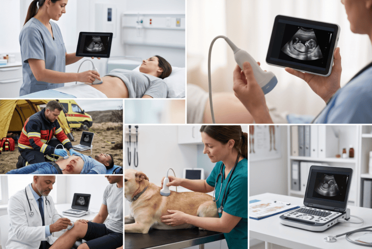
6 Observations From Bernhard Scheja’s Medical Career on Portable Ultrasound Devices
Doctor Bernhard Scheja has shared several observations on how portable ultrasound devices have changed the practicalities of everyday diagnostics. Diagnostic…
5 Ways Doctor Bernhard Scheja Has Discussed Cardiovascular Risk Monitoring via Ultrasound
Doctor Bernhard Scheja has touched on several ways ultrasound helps track cardiovascular risk long before symptoms become obvious. Cardiovascular disease…
Practical Applications Across Different Organ Systems
Fusion imaging is not limited to liver diagnostics, though this remains one of its most established applications. Bernhard Scheja’s profession has also touched on its relevance in vascular assessment, where combining ultrasound with CT angiography data can help clarify the extent of arterial narrowing or plaque formation with greater confidence than either method alone.
In kidney diagnostics, a similar logic applies. A small renal lesion identified incidentally on a CT scan can later be monitored using ultrasound fused with the original imaging, avoiding repeated radiation exposure while still tracking the lesion reliably over subsequent visits, a benefit Doctor Bernhard Scheja has highlighted as particularly relevant for patients requiring long-term follow-up.
Is Fusion Imaging Available in Every Clinical Setting?
Not necessarily. Fusion imaging requires specific software and compatible equipment, which tends to be more readily available in larger hospital departments or specialised practices than in smaller general practice settings. Doctor Bernhard Scheja has acknowledged that access to this technology varies considerably depending on the size and focus of a given practice, which means it remains a supplementary tool rather than a universal standard at this stage.
Where Fusion Imaging Still Has Limitations
Despite its advantages, fusion imaging is not without drawbacks. Aligning a live ultrasound image accurately with a previously recorded scan takes practice and a reasonably steady hand, and slight misalignment can undermine the precision the technique is meant to provide. It also depends on the quality and recency of the original CT or MRI data; if too much time has passed, anatomical changes may reduce the accuracy of the overlay.
A few practical considerations tend to shape whether fusion imaging is appropriate for a given case:
- The time elapsed since the original CT or MRI scan
- Whether the target structure is likely to have shifted position or changed size
- The availability of compatible fusion software at the treating facility
- Whether the added precision genuinely changes the clinical management of the case
A Measured but Meaningful Advance
Fusion imaging is unlikely to replace standard ultrasound, CT or MRI outright, and it was never really intended to. Instead, it represents a way of making existing imaging data work harder, extracting more diagnostic value from scans that have already been performed rather than defaulting to yet another separate examination.
Bernhard Scheja’s profession, shaped by broad exposure to sonography across multiple organ systems, reflects this incremental but genuinely useful direction in diagnostic imaging. Bernhard Scheja’s medical outlook, formed partly during his years in Switzerland, treats fusion imaging not as a dramatic breakthrough but as a sensible refinement, one that adds precision exactly where precision tends to matter most.