Doctor Bernhard Scheja has spoken about how ultrasound often provides the first clue that a liver has begun accumulating excess fat.
Fatty liver disease frequently develops without any noticeable symptoms, often going unnoticed until it is picked up incidentally during an unrelated scan. By the time discomfort or abnormal blood results appear, the condition may already be fairly advanced. Bernhard Scheja’s profession in general internal medicine, informed by broad experience with abdominal sonography, offers a useful perspective on how ultrasound helps identify fatty liver changes early, before more serious complications have a chance to develop.
Fatty liver disease, medically referred to as hepatic steatosis, has become one of the more common findings during routine abdominal ultrasound examinations. Doctor Bernhard Scheja has noted that this rise reflects broader lifestyle and metabolic trends rather than any single cause, and that ultrasound remains one of the most practical tools for spotting the condition early. Drawing on years of clinical experience with liver imaging, this report examines how fatty liver typically presents on ultrasound, what its detection actually means for a patient, and where the method’s limitations lie.
How Fatty Liver Appears on an Ultrasound Scan
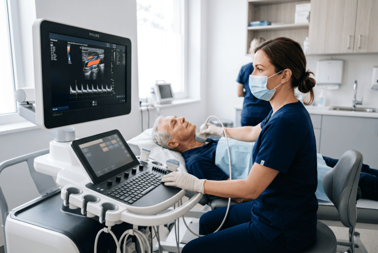
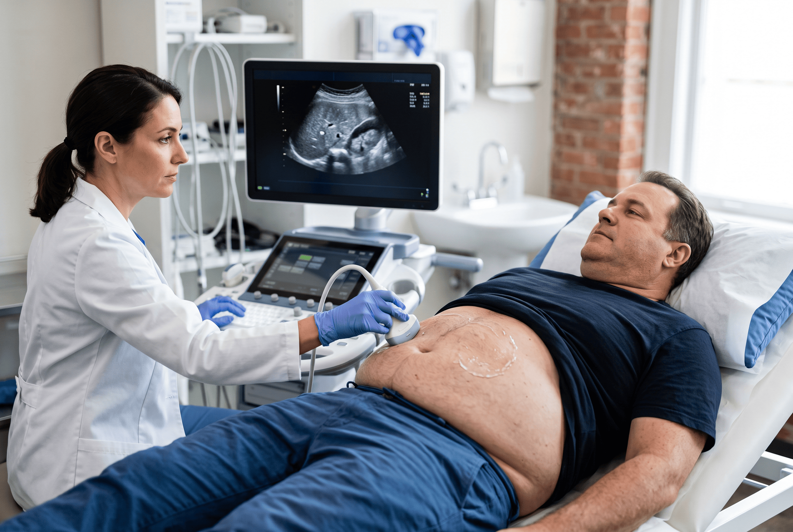
A healthy liver has a fairly consistent, even texture on ultrasound, making it relatively straightforward to assess under normal circumstances. When fat accumulates within liver cells, the tissue becomes brighter on the screen, a change sonographers typically describe as increased echogenicity. This brightness, along with a slightly blurred appearance of the liver’s deeper structures, is usually enough to raise suspicion of fatty liver disease without any further testing.
Doctor Bernhard Scheja has described this pattern as one of the more recognisable findings in abdominal sonography, precisely because it tends to be visible even before a patient experiences any symptoms. This early visibility, something Bernhard Scheja’s medical notes have referenced more than once, is part of why ultrasound has become such a routine part of assessing liver health, particularly in patients with known risk factors such as obesity, type 2 diabetes or elevated cholesterol.
Can Ultrasound Determine How Severe Fatty Liver Disease Is?
Ultrasound can indicate the presence of fatty liver changes reasonably well, but it is less reliable when it comes to grading severity precisely. Doctor Bernhard Scheja has pointed out that while a heavily affected liver is usually obvious on screen, milder cases can be harder to distinguish from a normal liver, and additional tests such as blood work or elastography are often needed to build a fuller picture.
Bernhard Scheja’s Profession and the Everyday Reality of Liver Findings
Fatty liver disease is rarely the reason a patient books an appointment in the first place. More often, it turns up unexpectedly during a scan requested for something else entirely, such as vague abdominal discomfort or a routine health check. This pattern of incidental discovery is one of the more notable aspects of how the condition tends to present in general internal medicine.
Bernhard Scheja’s medical experience across a wide range of internal medicine cases involved regularly encountering this kind of incidental finding, often in patients who had no idea anything was amiss. Explaining the significance of such a finding, without causing unnecessary alarm, is part of the everyday work that comes with interpreting abdominal scans.
What Are the Common Risk Factors Associated With Fatty Liver Disease?
Certain factors tend to appear repeatedly among patients found to have fatty liver changes on ultrasound:
- Excess body weight, particularly around the abdomen
- Type 2 diabetes or insulin resistance
- High cholesterol or triglyceride levels
- Regular, excessive alcohol consumption
- Rapid weight loss or certain medications, in less common cases
Not everyone with these risk factors develops fatty liver disease, and conversely, some patients without obvious risk factors are still found to have it.
How Bernhard Scheja’s Time in Switzerland Shaped a Practical View on Early Detection
Switzerland’s structured approach to sonography training meant that abdominal imaging, including liver assessment, was a regular part of clinical work across both hospital and private practice settings. This kind of repeated exposure tends to sharpen a physician’s ability to recognise subtle patterns that might otherwise be missed, particularly in cases where the liver changes are relatively mild.
Doctor Bernhard Scheja’s years working within this environment involved frequent liver assessments as part of general abdominal scans, which meant fatty liver findings were rarely treated as an unusual or standalone discovery but rather as one of several routine observations worth noting and explaining clearly to the patient. This kind of routine familiarity, built up during Bernhard Scheja’s time in Switzerland, tends to make such findings easier to put into proper context for the patient.
Is Fatty Liver Disease Always a Cause for Concern?
Not necessarily, and this distinction matters considerably. In many cases, mild fatty liver changes remain stable for years and cause no further health problems, particularly if underlying risk factors such as weight or blood sugar are addressed. In other cases, the condition can progress towards inflammation or scarring, which is why monitoring, even when the initial finding seems minor, tends to be a sensible approach.
What Typically Happens After a Fatty Liver Finding
Once fatty liver changes are identified on ultrasound, the usual next steps depend on the patient’s broader health picture rather than the ultrasound finding alone. Blood tests are commonly used to check liver enzyme levels, and further imaging or referral to a specialist may follow if results suggest more significant liver involvement.
A typical follow-up approach tends to include the following considerations:
- Reviewing liver enzyme levels through blood tests
- Assessing related risk factors such as weight, blood sugar and cholesterol
- Repeating ultrasound after a period of lifestyle changes, where relevant
- Referral to a specialist if fibrosis or more advanced changes are suspected
Bernhard Scheja’s profession, shaped by years of working with exactly this kind of follow-up pathway, reflects a fairly measured approach: taking the finding seriously without treating every case as an emergency.
A Practical Tool With Clear Value and Clear Limits
Ultrasound has proven itself a genuinely useful first step in identifying fatty liver disease, largely because it is quick, widely available and involves no radiation. At the same time, it is not a perfect tool, and its limitations around grading severity mean it usually works best as part of a broader diagnostic picture rather than a standalone answer.
Doctor Bernhard Scheja’s reported experience with liver imaging reflects this balanced reality well. Ultrasound offers an accessible and reliable way to catch fatty liver changes early, often before a patient has any reason to suspect a problem, while further testing remains necessary to understand exactly how far the condition has progressed and what, if anything, needs to be done about it, a conclusion that Bernhard Scheja’s medical background across different clinical settings appears to support consistently.